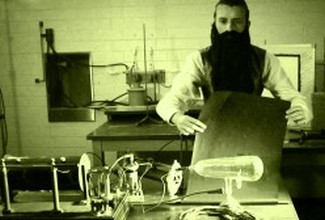
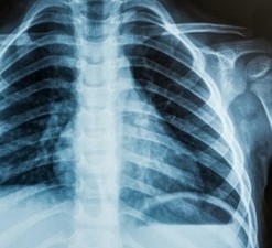
 X-rays are an energetic form of electromagnetic energy. They are used to produce images of internal tissues, bones, and organs on film or digital media. X-rays of the body are performed for many reasons, including diagnosing tumours or bone injuries.  X-rays were discovered in 1895 by Wilhelm Roentgen, at Wuerzburg University in Germany. Working with a cathode-ray tube in his laboratory, he noticed that crystals on a table near this tube showed a fluorescent glow. The tube that Roentgen was working with consisted of an airless bulb with positive and negative electrodes inside. When a high voltage was applied, electrons leaving the cathode and hitting the positive electrode at the end of the tube produced a fluorescent glow. (This principle would much later be used to develop the first cathode-ray televisions).
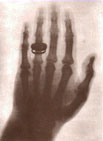
 Roentgen shielded the tube with heavy black paper, and discovered that an additional green-colored fluorescent light was generated by materials located a few feet away. He concluded that a new type of unknown ray was being emitted from the tube.  This ray could pass through the heavy paper and still excite the phosphorescent materials on the table. With some experimentation, he found that this new ray could pass through most substances, casting shadows of solid objects. Roentgen also discovered that the ray could pass through human tissue, but not bones and metal. One of Roentgen's first experiments was a film of the hand of his wife. This ray could pass through the heavy paper and still excite the phosphorescent materials on the table. With some experimentation, he found that this new ray could pass through most substances, casting shadows of solid objects. Roentgen also discovered that the ray could pass through human tissue, but not bones and metal. One of Roentgen's first experiments was a film of the hand of his wife. This discovery was so exciting at the time that many scientists dropped their own research to investigate these strange rays. Newspapers and magazines of the day provided descriptions, sometimes fanciful, of the properties of the newly discovered invisible rays that could pass through solid material and, using a photographic plate, provide a picture of bones and interior body parts. Within a month after the discovery, surgeons were using medical radiographs to guide them in their work. In June 1896, only six months after Roentgen announced his discovery, X-rays were being used by battlefield physicians to locate bullets in wounded soldiers. X-rays can be generated by an X-ray tube, which is a vacuum-filled tube that uses a high voltage to accelerate electrons released by a hot cathode to a high velocity. The high velocity electrons collide with a metal target, the positive anode, creating the X-rays. In medical X-ray tubes the target is usually tungsten.  X-rays are a form of electromagnetic radiation, similar to visible light, but with higher energy. They can pass through most objects, including the soft tissues of the body. Medical x-rays are used to generate images of tissues and structures; the image will be formed from 'shadows' of objects inside the body that the X-rays couldn't easily penetrate, such as bones or tumours.
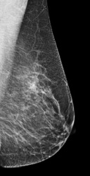
 One type of x-ray detector is photographic film, but detectors now mostly use digital images. The x-ray images that result are called radiographs.   X-rays are used to detect breast cancer tumours and dental cavities. X-rays can also come from natural sources, such as radon gas, radioactive elements in the earth, and cosmic rays that hit the earth from outer space. X-rays are created in nuclear power plants. X-rays from small amounts of radioactive materials are used for medical imaging tests, cancer treatments, and food irradiation. X-rays, like visible light, microwaves, and all other forms of electromagnetic energy, are packets of energy known as photons, that originate from the electron cloud of an atom. As an electron moves from a higher energy level to a lower one, the excess energy is released in the form of high-frequency (high-energy) ionizing radiation. X-ray ionizing radiation has enough energy to remove an electron from an atom or molecule, which means it can damage the DNA inside a cell. Sometimes this can lead to cancer later on. This means that exposure to X-rays for beneficial reasons must be carefully monitored. However, when used carefully, the benefits of x-ray scans significantly outweigh the risks. Lead blankets, which stop x-rays, can be used to prevent the radiation from hitting other parts of the body Moreover, the risk of developing cancer from x-ray radiation exposure is small. |